Thermography or Mammography?
This is one of the first questions ladies ask when they hear about Breast Thermography, and there is a simple answer to it: they are two completely different procedures with two different targets. Can one replace the other? No, each serves a different purpose.
Mammography looks for EXISTING masses or tumors by compressing breast tissue and sending X-rays through it. The tumor has to be large enough (generally several years in development) and the breast tissue shouldn’t be dense (as it is the case in younger ladies).
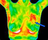
Thermography looks for the process of a tumor build up. From very early days of abnormal cell multiplication, new blood vessels will be made to nourish them and so the temperatures in that area rises. Thermography camera registers this temperature rise thereby giving a very early warning signal that the area must be monitored and necessary steps(such as dietary changes etc.) taken. The breast surface temperature reading is done with an infrared camera from several feet away without touching the breast and without sending any radiations to it. The target in Thermography procedure is not to detect a large tumor but to detect small abnormalities in the breast tissue so that the person will take preventive measures to not allow a large tumor build up. Does thermography also see large abnormalities in breast (such as a large tumor)? Absolutely yes. When the camera can see the temperature rise due to a few hundred new cells, it clearly shows the existence of millions of new (tumor) cells. Thermography can be used for any gender and age group.
Neither Mammography nor Thermography can tell if the cell increase(tumor) is cancerous! Only a biopsy (removing some of breast tissue and analyzing it) can determine this. The objective in Thermography is not let a tumor development go unnoticed for many years till it is detectable, then going through the biopsy process or in an unlucky case deal with the already spread of a cancerous tumor to other parts of the body, rather giving an early warning to take action and prevent all that complications.
Video: Mammogram and Thermography
Video: Dr. Mercola on Breast Thermography
Video: Preventing Breast Cancer
Video: News on Thermography on CBS
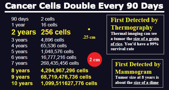
Cancer Cell Multiplication
Indications for a Thermographic Evaluation
| Altered Ambulatory Kinetics | Nerve Entrapment |
| Altered Biokinetics | Nerve Impingement |
| Arteriosclerosis | Nerve Pressure |
| Brachial Plexus Injury | Nerve Root Irritation |
| Biomechanical Impropriety | Nerve Stretch Injury |
| Breast Disease | Nerve Trauma |
| Bursitis | Neuropathy |
| Carpal Tunnel Syndrome | Neurovascular Compression |
| Causalgia | Neuralgia |
| Compartment Syndromes | Neuritis |
| Cord Pain/Injury | Neuropraxia |
| Deep Vascular Disease | Neoplasia |
| Disc Disease | (melanoma, squamous cell, basal) |
| Disc Syndromes | Nutritional Disease |
| Dystrophy | (Alcoholism, Diabetes) |
| External Carotid Insufficiency | Peripheral Nerve Injury |
| Facet Syndromes | Peripheral Axon Disease |
| Grafts | Raynaud’s |
| Hysteria | Referred Pain Syndrome |
| Headache Evaluation | Reflex Sympathetic Dystrophy |
| Herniated Disc | Ruptured Disc |
| Herniated Nucleus Pulposis | Somatization disorders |
| Hyperaesthesia | Soft Tissue Injury |
| Hyperextension Injury | Sprain/Strain |
| Hyperflexion Injury | Stroke Screening |
| Inflammatory Disease | Synovitis |
| Internal Carotid Insufficiency | Sensory Loss |
| Infectious Disease (Shingles, | Sensory Nerve Abnormality |
| Leprosy) | Somatic Abnormality |
| Lumbosacral Plexus Injury | Superficial Vascular Disease |
| Ligament Tear | Skin Abnormalities |
| Lower Motor Neuron Disease | Thoracic Outlet Syndrome |
| Lupus | Temporal Arteritis |
| Malingering | Trigeminal Neuralgia |
| Median Nerve Neuropathy | Trigger Points |
| Morton’s Neuroma | TMJ Dysfunction |
| Myofascial Irritation | Tendonitis |
| Muscle Tear | Ulnar Nerve Entrapment |
| Musculoligamentous Spasm | Whiplash |
More Thermography Applications
- Infections
- Skin cancer
- Neuropathies
- Superficial vascular disorder
- Musculoskeletal injuries
- Whiplash
- Headache/Migraine
- Chronic Fatigue Syndrome
- Pre-Stroke
- Heart disorders
- Periodontal disorders/TMJ
- Muscular Inflammation
- Diverticulitis
- Fibromyalgia
- Carpal Tunnel Syndrome
- Vericosities
- Scoliosis
- Cystic Formations
- Pre-Heart Attack
- Thyroid Dysfunction
- Inflammatory pain
- Cardio-Vascular Inflammation
- Digestive disorders
- Immune System Response
- And much more…
Evaluating the Thermography Images
The Thermography images taken in our office are “read” by either Dr. Ben Johnson or Dr. Greg Melvin. Dr. Gregory Melvin, DC, BCCT, is a Board Certified Clinical Thermographer with 20 years experience in the field of Medical Thermography. As the published author of “The Secret of Health: Breast Wisdom,” Dr. Ben Johnson, MD, DO, NMD understands the significance of thermography and contributes a chapter of his book to the subject of thermography.
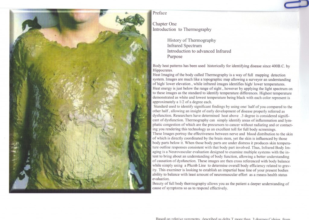
Dr. Johnson About Thermography
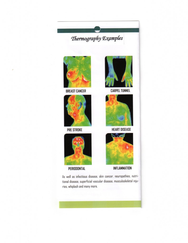
Thermography Examples
A Few Facts About DITI
- Medical thermography was approved by USFDA in 1982 as an adjunctive procedure for breast cancer risk assessment.
- DITI is specially useful for women with dense breast tissue and women with surgically altered breast.
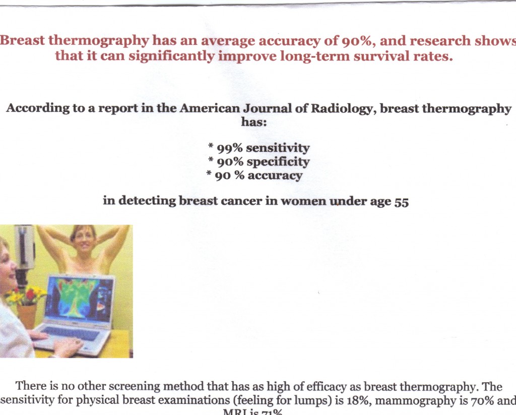
- For people under 55 years old, DITI has a 99% sensitivity, a 90% specificity and 90% accuracy.
- DITI is used for DCIS (Ductal Carcinoma In Situ), where cancer cells grow in ducts and so develop without forming a palpable mass!
- DITI detects inflammation, a precursor for many disorders.
- DITI detects irregularities in breast YEARS before a lump is formed.
- DITI is an excellent breast cancer monitoring/assessment tool for women of even young ages.
- Thermography is the only method that can “Visualize” pain, being acute or chronic.
- DITI is a good warning tool for stroke and blood clots by detecting inflammation in carotid arteries.
- DITI can differentiate between osteoarthritis and rheumatoid arthritis. It is also a good warning tool for early assessment of arthritis.
- DITI may point to immune system problems by detecting heat at specific vertebrae.
What Is Thermography
What is Thermography?
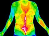
Thermography — or more accurately said for our purpose — Digital Infrared Thermal Imaging (DITI), is an industry technique to capture remotely objects’ surface temperatures. Most of us probably have seen this method at least in the movies when somebody holds a “binocular” and “sees” people in the darkness of the night. Those binocular-looking devices are nothing but an Infrared Camera that detects different surface temperatures of objects (being a human, an animal or any other object). Objects in nature are exposed to sun infrared radiation and emit themselves at different rates the infrared energy. The “Infrared Cameras” read with a high degree of accuracy these emitted infrared temperatures off of the surface of an object and the read temperatures can be plotted (with the help of a computer) to “see” the whole or part of the object.
The human body constantly emits infrared energy from the skin surface. The skin is in “communication” with every part of our body through our Nervous System. The “abnormalities” of the body translate into different skin temperatures.
Let us take the “breast thermography” as an example. The infrared camera takes a picture from your breast area (from a few feet away). The camera is capable of reading the temperatures of hundreds of single points of the breast surface. In case of a developing tumor (even in the very earliest stages) the skin temperature will rise as the new tumor cells build their “own” blood vessels, needed for their nourishment, causing a rise of temperature on the skin (via Sympathetic Nervous System). This simple process is so nothing more than taking a photograph with an infrared camera, without a contact to the body and without “sending” anything to it and can be used to monitor the breast (or other parts of the body) of women and men at any age group with zero side effects.
The thermography principle is based on the fact that our Sympathetic Nervous System responds to “physiological” changes anywhere in the body. Unlike other screening methods that target “structural” changes — such as formation of a tumor which has to be large enough to be detected, requiring several years up to a decade — thermography targets the “path” the tumor has to go from a few cells to a large detectable tumor, i.e. the physiological changes in the body that appear parallel to the abnormality formation.
Sensitivity and Accuracy
Thermography Principle in Ancient History
Thermography Screening Fees
Breast and Lymph
|
$249.00 |
Complete Women’s Health
|
$395.00 |
| Women Full Body: From Head to Toe | $449.00 |
| Complete Men’s Health | $349.00 |
| Men Full Body: From Head to Toe | $399.00 |
| Region of Interest (Male or Female) | $229.00 |
Excerpts from Summary of Studies
The Biomedical Handbook »»
“1982 FDA approved Thermography as an adjunctive Breast screening procedure. Breast Thermography has the ability to detect the first signs of a tumor that may be forming up to 10 years before other procedures can detect it.”
National Cancer Institute »»
“Thermography’s digital infrared imaging can detect the increase in regional breast temperatures resulting from measured blood vessel activity in both pre cancerous tissue and the area surrounding a developing breast cancer.”
The Lancet »»
“Screening for breast cancer with mammography is unjustified…the data show that for every 1000 women screened biennially throughout 12 years, one breast-cancer death is avoided whereas the total number of deaths is increased by six…there is no reliable evidence that screening decreases breast-cancer mortality.”
2003 issue of AMA journal, American Medical News »»
“There is little evidence documenting that mammography saves lives from breast cancer for premenopausal women.”
THERMAL IMAGING CAN DETECT MANY DISEASES AND DISORDERS IN THEIR EARLY STAGES
Thermography is a non-invasive imaging technique
Thermography uses special infrared-sensitive cameras to digitally record images of the variations in surface temperature of the human body. It’s basically a specialized HDTV night vision camera used to record images of the heat patterns of the breast. The recorded images are called thermograms.
Thermography is an unsurpassed, safe and noninvasive breast cancer screening technique that can detect signs of cancer up to ten years earlier than is possible using mammography. Breast thermography is approved by the FDA for breast cancer risk assessment. The exam takes only a few minutes, and there is no touching or compression of the breast whatsoever.

